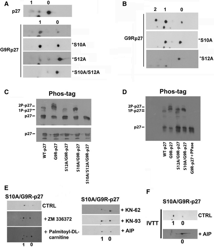
Fig. 3.

Characterization of G9R‐p27 phosphorylation. (A) pcDNA3.0 plasmids encoding p27, G9R‐p27, S10A/G9R‐p27 (*S10A), S12A/G9R‐p27 (*S12A), and S10A/S12A/G9R‐p27 (*S10A/S12A) were translated and transcribed in vitro as reported under Materials and methods. Each IVTT assay mixture was then analyzed by 2D/WB as reported in Figure 2. The filters were analyzed by anti‐p27 mAb. Signals 0 and 1 correspond to unmodified and 1Pi‐proteins, respectively. (B) MEF cells were transfected with three different pcDNA3.0 plasmids encoding G9R‐p27, S10A/G9R‐p27 (*S10A), and S12A/G9R‐p27 (*S12A). After 24‐h transfection, cells were pelleted and cell extracts were prepared as reported under Materials and methods. Equal amounts of protein were analyzed by 2D/WB employing anti‐p27 mAb. Signals 0, 1, and 2 correspond to unmodified, 1Pi‐, and 2Pi‐proteins, respectively. (C) PC‐3 cells were transfected with five different pcDNA3.0 plasmids encoding WT‐p27, G9R‐p27, S12A/G9R‐p27, S10A/G9R‐p27, and S10A/S12A/G9R‐p27. After 24‐h transfection, cells were pelleted and extracts were prepared as reported under Materials and methods. Then, equal amounts of protein were analyzed by 1D/WB (lower image) and SDS/PAGE on polyacrylamide gel strengthened with agarose containing 20 mm Mn2+–Phos‐tag (upper image). The filters were analyzed by anti‐p27 mAb. Further details are reported under Materials and methods Section. (D) An experiment similar to that reported in Panel C is showed, except that an aliquot of extract from PC‐3 cells expressing G9R‐p27 (prepared without phosphatase inhibitors) was treated with protein phosphatase lambda (PPase). The samples were then analyzed by Phos‐tag SDS/PAGE, as reported in panel C and in Materials and methods Section. (E) PC‐3 cells were transfected with pcDNA3.0 encoding S10A/G9R‐p27 in order to study only the phosphorylation of G9R‐p27 on S12. Then, cRaf inhibitor (ZN336372), PKC inhibitor (palmitoyl‐DL‐carnitine), and three CAMK II inhibitors (KN‐62, KN‐93, and AIP) were added 2 h before the transfections. After 24 h, cell extracts were prepared and analyzed by 2D/WB employing anti‐p27 mAb compared with cells treated with vehicle as CTRL. The employed concentrations of the compounds are reported in Table 1. (F) pcDNA3.0 encoding S10A/G9R‐p27 plasmid was transcribed and translated in vitro in the absence (CTRL) and presence of AIP (CAMKII inhibitor). The mixtures were analyzed by 2D/WB employing anti‐p27 mAb.
