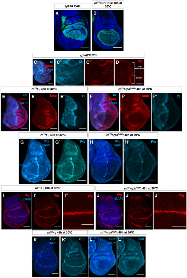
Figure EV4. Manipulating levels of the ion channel Rpk impacts both Hh and Wg signal transduction.

-
A, BExpression patterns of ap‐Gal4 (A) and rn‐Gal4 (B).
-
C, DImmunostaining of Smo protein (red), full‐length Ci (light blue) (C–C″) and Ptc protein (D) in discs expressing ATPαRNAi in the dorsal compartment. Brackets indicate tissue expressing RNAi and control tissue.
-
E–F″Immunostaining of Smo protein (red) and full‐length Ci (light blue) in (E–E″) control discs and (F–F″) discs expressing rpkRNAi for 48 h before dissection.
-
G–H′Immunostaining of Ptc protein in (G, G′) control discs and (H, H′) discs expressing rpkRNAi for 48 h before dissection.
-
I–J″Immunostaining for Wg protein in (I–I″) control discs and (J–J″) discs expressing rpkRNAi for 48 h before dissection.
-
K–L′Immunostaining for Cut protein in (K, K′) control discs and (L, L′), discs expressing rpkRNAi for 48 h before dissection.
Data information: All scale bars are 100 µm except (D), where scale bar is 50 µm.
