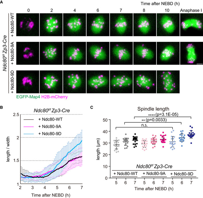
Figure 4. Kinetochore–microtubule attachment stability is not required for initiating but is required for terminating spindle elongation.

- Live imaging of Zp3‐Cre Ndc80f/f oocytes injected with Ndc80‐WT, Ndc80‐9D or Ndc80‐9A. Z‐projection and 3D‐reconstructed images show microtubules (EGFP‐Map4, green) and chromosomes (H2B‐mCherry, magenta). Scale bar, 10 μm.
- Ndc80‐9D is defective for limiting spindle elongation. The aspect ratio (length/width) of 3D‐reconstructed spindles was measured over time (n = 18, 18, and 18 oocytes, respectively, from three independent experiments).
- Ndc80‐9D causes excessive spindle elongation. Spindle lengths measured after 3D reconstruction are shown (n = 18, 18, and 18 oocytes, respectively, from three independent experiments). Two‐tailed Student’s t‐test was performed. **P < 0.01, ****P < 0.0001.
Data information: Means ± SD are shown. See also Movie EV8.
Source data are available online for this figure.
