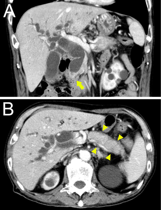
Figure 1.

CT images revealing a low-density mass lesion measuring 15 mm in diameter at the pancreatic head (orange arrow) with bile duct dilation proximal to the mass lesion (A) and diffuse enlargement and a surrounding low-density line of the pancreatic body and tail that was considered a capsule-like rim (B).
