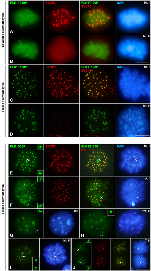
Figure EV2. Distribution of PLK1S137P and PLK1T210P.

-
A, BDouble immunolabelling of PLK1T210P (green) and SYCP3 (red) on squashed spermatocytes at (A) metaphase I and (B) metaphase II. Chromatin has been stained using DAPI (blue).
-
C, DDouble immunolabelling of PLK1T210P (green) and SYCP3 (red) on spread spermatocytes at (C) metaphase I and (D) metaphase II. Chromatin has been stained using DAPI (blue).
-
E–JDouble immunolabelling of PLK1S137P (green) and SYCP3 (red) on spread spermatocytes at (E) metaphase I, (F) anaphase II, (G) interkinesis, (H) prophase II, (I) metaphase II and (J) telophase II. Chromatin has been stained using DAPI (blue). Insets correspond to centrosomes at 200% magnification.
Data information: White arrows indicate the location of centrosomes. Scale bar in D and J represents 10 μm.
