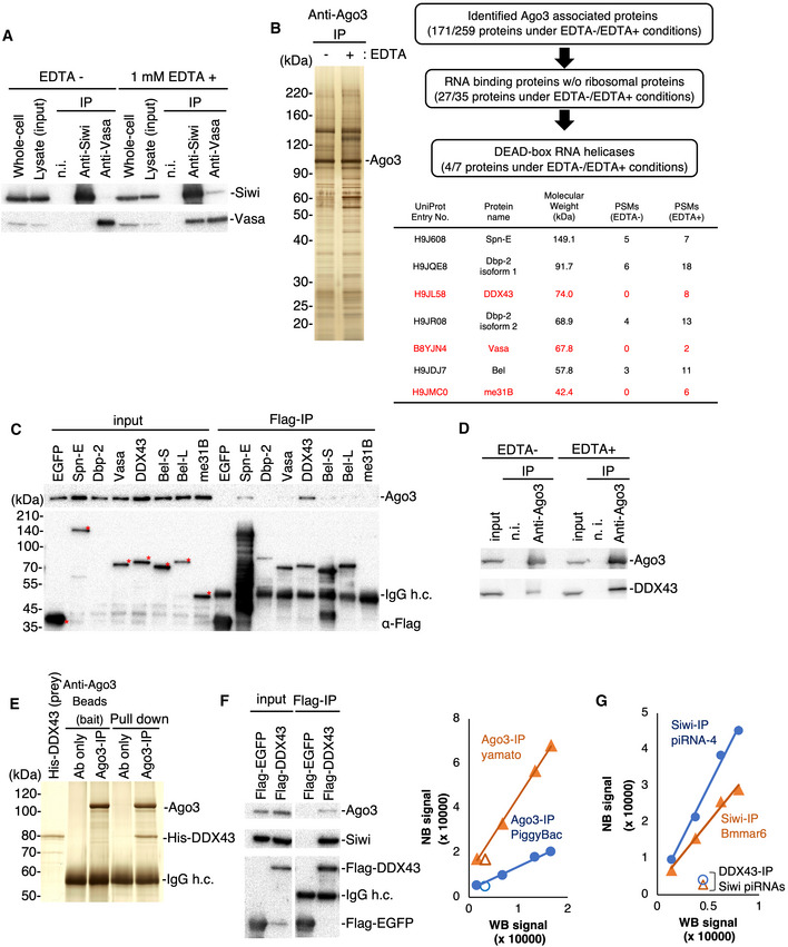
Figure 1. Identification of RNA helicase DDX43 associated with Ago3.

-
ACo‐immunoprecipitation of the Siwi–Vasa complex in the presence and absence of EDTA. Isolated proteins were subjected to Western blotting. n.i.: non‐immune mouse IgG.
-
BCo‐immunoprecipitation and identification of Ago3‐associated proteins. Left: The immunopurified Ago3 complex was resolved by SDS–PAGE and silver‐stained. Right: DEAD‐box helicases identified by mass spectrometry. Proteins identified by co‐immunoprecipitation under EDTA‐containing conditions only are listed in red. PSM: peptide sequence match.
-
CCo‐immunoprecipitation using BmN4 cells transfected with the Flag‐tagged DEAD‐box helicases identified by shotgun proteomics. The Flag‐tagged proteins and Ago3 were detected by Western blotting. Full‐length Flag‐tagged proteins are indicated with red asterisks. h.c.: heavy chain.
-
DCo‐immunoprecipitation of endogenous Ago3 and DDX43 in the presence or absence of EDTA. These factors were detected by Western blotting.
-
EPull‐down assay using high salt‐washed Ago3‐conjugated magnetic beads and purified His‐DDX43. All samples were subjected to SDS–PAGE and detected by silver staining.
-
F, GAnalysis of Siwi‐piRNA and Ago3‐piRNA levels in Siwi and Ago3 complexes with DDX43, respectively. The WB (Western blotting) and NB (Northern blotting) signals represent the PIWI protein and piRNA levels, respectively (see also Fig EV1A and B). The relative ratios of piRISC were calculated based on the signal intensities of the PIWI proteins and piRNAs.
