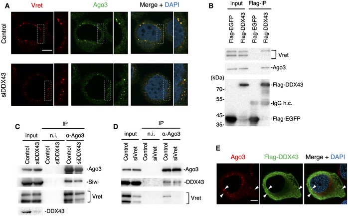
Figure 2. DDX43 depletion does not affect the formation of the Ago3–Vret complex.

-
AImmunofluorescence showing the Vret (red) and Ago3 (green) signals in DDX43‐depleted BmN4 cells (siDDX43). DAPI shows the nuclei (blue). Scale bar: 10 µm. The insets show high‐magnification images of the part indicated sections.
-
BImmunoprecipitation and Western blotting showing that DDX43 associated with Vret and Ago3.
-
C, DImmunoprecipitation of endogenous Siwi, DDX43, and Vret bound with Ago3 from DDX43‐depleted (C) or Vret‐depleted (D) cells. The factors were detected by Western blotting. Ago3 interacts with Vret and DDX43 independently of the other.
-
EImmunofluorescence showing Ago3 (red) and Flag‐DDX43 (green) signals in BmN4 cells. DAPI shows the nuclei (blue). Arrowheads indicate colocalization of these factors. Scale bar: 10 µm.
