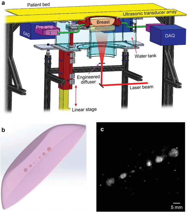
Figure 1.

Schematic of the SBH‐PACT and breast phantom imaging. a) Perspective cut‐away view of the SBH‐PACT breast imaging system. DAQ, data acquisition module; Pre‐amp, preamplifiers. b) Sketch of the breast‐mimicking phantom. c) Maximum‐amplitude‐projection (MAP) of the breast phantom image acquired by SBH‐PACT, which revealed the smallest tumor phantom (1‐mm diameter). The bright dots in the image background were caused by air bubbles embedded in the phantom.
