
Figure 3.
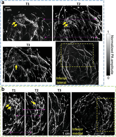
SBH‐PACT images of the other two patients treated with NAC. a) Angiograms of Patient 2 who had a partial response to NAC. Close‐up views of the cancer‐affected area are shown from T1 to T3 in the region outlined by yellow dashed boxes (bottom right corner of figure). Breast cancer is identified by yellow arrows. Prominent common blood vessels in T1 and T2 images are marked by magenta arrow lines. b) Angiograms of Patient 3 who had a complete clinical response to NAC. Angiogenesis associated with the cancer was not detected at T3.