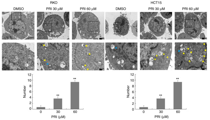
Figure 4.
Autophagosomes and autolysosomes are detected via transmission electron microscopy (TEM) after treatment with DMSO, 30 µM PRI or 60 µM PRI for 24 h (blue arrow, autophagosome; yellow arrow, autolysosome). Increased numbers of autophagosomes and autolysosomes were observed in RKO and HCT15 cells treated with PRI. Data were obtained from three different cells in each treated sample and presented as the mean + SEM. **P<0.01, compared with the PRI untreated cells. PRI, p53-reactivation and induction of massive apoptosis-1, APR-017 methylated.

