Figure S3.
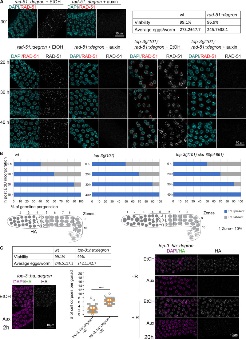
In top-3, meiotic progression is not delayed. Related to Fig. 4 and Fig. 5. (A) Top: representative rad-51::degron nuclei in pachynema stained with DAPI (cyan) and anti-RAD-51 (red) exposed to ethanol (EtOH) or auxin for 30 min, time needed for RAD-51 depletion. Hatch rates and brood sizes of rad-51::degron (n = 15 worms) and wt (n = 12 worms). Bottom: representative rad-51::degron and top-3(jf101); rad-51::degron nuclei in pachynema stained with DAPI (cyan) and anti-RAD-51 (red) exposed to ethanol (EtOH) or auxin at different time points. (B) Top: The charts indicate EdU incorporation in the germline, divided into 10 equal zones. EdU incorporation was quantified: EdU present (blue) or EdU absent (gray). For the indicated genotypes, four time points were analyzed. Bottom: Schematic representation of wt and mutant gonads divided into 10 equal zones to show the prolonged mitotic zone in top-3. (C) Left: Functionality of the top-3::ha::degron strain. Hatch rates and brood sizes of top-3::ha::degron (n = 23 worms); and wt (n = 10 worms). Representative top-3::ha::degron nuclei in pachynema, stained with DAPI (magenta) and anti-HA (green) exposed to EtOH or auxin for 2 h, time needed for TOP-3 depletion. Apoptosis quantification of top-3::ha::degron with and without irradiation with SYTO-12. Scatter plots indicate the mean ± SD. n = number of gonads scored: top-3::ha::degron not irradiated, 2.4 ± 1.5, n = 72; top-3::ha::degron after irradiation, 6.2 ± 1.5, n = 48. ****, P < 0.0001, calculated using the Mann–Whitney test. Right: Representative top-3::ha::degron nuclei in pachynema, stained with DAPI (magenta) and anti-HA (green) exposed to EtOH or auxin with and without irradiation (120 Gy). Aux, auxin; wt, wild-type.

