
Figure 1.
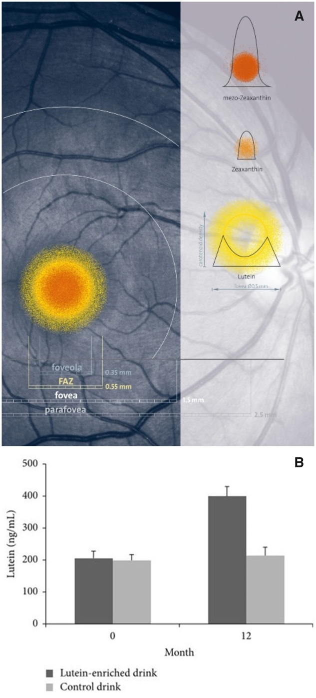
(A) Distribution of macular pigments lutein, meso-zeaxanthin, and zeaxanthin, presented in scale in a photograph of a healthy human retina. Image from Robert Kochling, Berlin, Germany; and John Nolan, Waterford, Ireland, with permission. (B) Mean ± standard error of plasma lutein concentration at 0 and 12 months for the lutein (dark grey) and the placebo (light grey) groups.207Abbreviation: FAZ, foveal avascular zone.