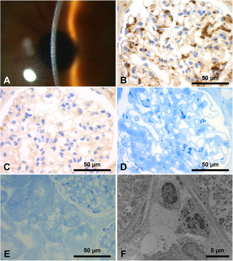
Fig. 1.

Slit lamp examination shows diffuse intracorneal crystalline deposits (a); immunohistochemistry staining for kappa light chains shows positivity of the crystals in the podocytes (b) with lambda negativity (c); glomerular staining with methylene-azure-blue stains light chain crystals strongly blue with a crystalline appearance (d); tubular staining with methylene-azure-blue shows scarce crystalline inclusions in the cytoplasm of proximal tubular epithelial cells (e); transmission electron microscopy confirms these crystalline inclusions diagnostic for minimal proximal light chain tubulopathy as intralysosomal (f)
