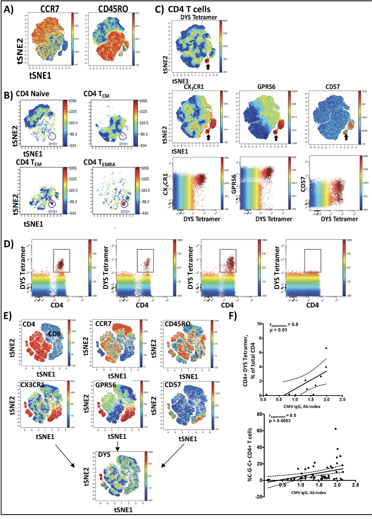
Figure 2. CMV-specific CD4+ T cells predominantly express the C~G~C surface marker combination.

PBMCs from 10 individuals with HLA-DR7 were stained with fluorescently tagged antibodies against surface marker proteins and the DYS tetramer. viSNE plots (n=10) were generated in Cytobank software and concatenated using flowjo software (A). DYS tetramer-positive CD4+ T cells were mainly TEM and TEMRA cells (B). DYS tetramer-positive cells (C, top panel) express CX3CR1, GPR56 and CD57 (i.e., ‘C~G~C cells’; C, middle panel). The Blue-Green-Yellow-Red gradient is the mean fluorescence intensity of DYS tetramer, with the surface markers on the right (C, middle panel). Two dimensional Flowjo plots showing CX3CR1and GPR56 expression on tetramer-positive and tetramer-negative CD4+ T cells (C, lower panel). DYS tetramer gating on total CD3+ T cells shows that it specifically binds to CD4+ T cells with no binding of CD8 T cells (D). Representative viSNE plot (n=1) of CD3+ T cells confirms that bright DYS tetramer+ cells bind CD4+ T cells and not CD8+ T cells (E). Correlation between CD4+ DYS tetramer-positive (n=10) and CD4+ C~G~C+ T cells with CMV IgG titers (n=57) (F). Correlations assessed by Spearman’s test, p<0.05 significant.
