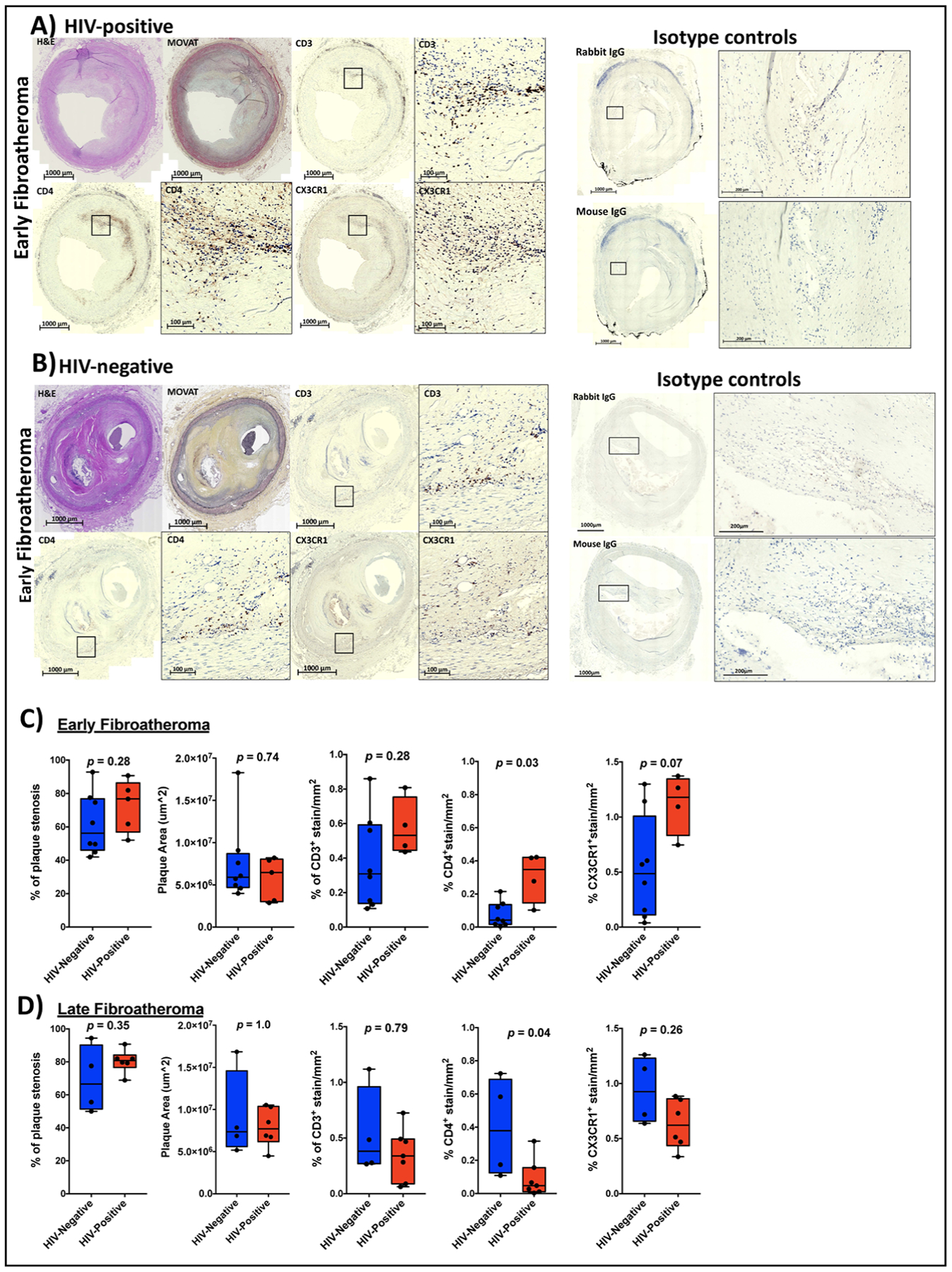
Figure 4. Immunohistochemical stain showing CX3CR1+ and CD4+ T cells within the coronary plaques of HIV-positive and HIV-negative persons.

Representative early atheroma images from one HIV-positive (A) and HIV-negative coronary plaque (B) stained with H&E stain, Movat stain, CD3, CD4 and CX3CR1. Box plots showing % stenosis, plaque area, % of CD3+, CD4+ and CX3CR1+ staining per mm2 coronary plaque area by HIV status in early atheroma (n=8 in HIV negative and n=5 HIV-positive, CD3+ stains on fewer samples; n=4 in HIV negative and n=6 HIV-positive) (C) and late atheroma (n=8 in HIV negative and n=5 HIV-positive) (D). Statistical analysis, Mann-Whitney test; p<0.05 was considered significant.
