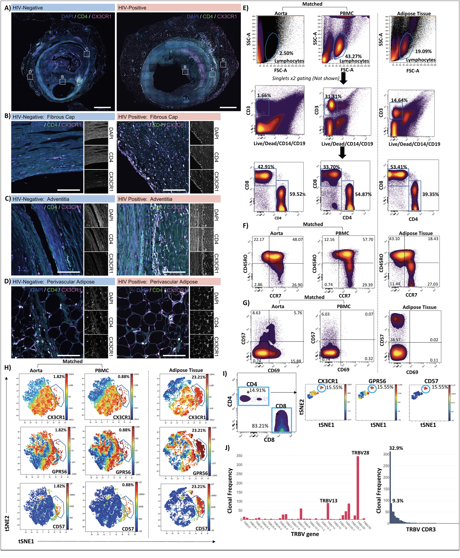
Figure 5. CX3CR1+ GPR56+ CD57+ CD4+ T cells are present in coronary arteries and aorta.

Low magnification confocal images of whole coronary plaques from HIV-negative (left) and HIV-positive (right) persons labeled with DAPI (blue), anti-CD4 (green), and anti-CX3CR1 (magenta), Scale bars 1000 μm (A). Enlarged images of the intima/ fibrous cap (B), adventitia (C) and perivascular adipose (D), scale bars 500 μm. Single cells isolated from the aorta of an HIV-negative individual were stained with the 12-panel antibodies including CX3CR1, GPR56 and CD57. PBMCs from the same person and subcutaneous adipose tissue from an HIV-positive participant as a positive control for tissue C-G-C+ CD4+ T cells were stained simultaneously. Lymphocytes, single cells, CD3+ live, CD4+ and CD8+ T cells were gated as shown (E). T cell memory populations were gated based on CCR7 and CD45RO co-expression (F). Two dimensional plots show expression of CD57 and CD69 (G). viSNE plots show C-G-C+ CD4+ T cells from aorta and PBMCs of HIV-negative donor and HIV-positive subcutaneous adipose tissue (H). C-G-C+ CD4+ T cells identified in a second HIV-negative aorta (I). Out of 949 CD3+ T cells sequenced from cells isolated from the aorta of donor 2; 240 clones were identified. The TCR β V gene and CDR3 clonal frequencies are shown (J).
