
Figure 4.
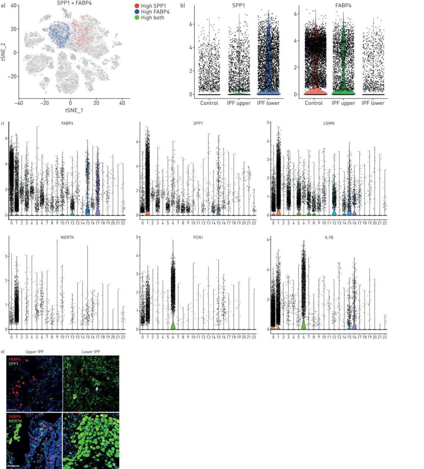
Macrophage populations in idiopathic pulmonary fibrosis (IPF) lungs. a) SPP1 and FABP4 expression define two discrete populations in IPF and normal lungs. b) (and supplementary figure S18) SPP1hi macrophages express more SPP1 and make up a higher percentage of cells in lower lobes, while FABP4hi macrophages are a higher percentage of the cells in healthy and IPF upper lobes. c) Violin plots of combined IPF/control lung data show expression of FABP4, SPP1, LGMN, MERTK, FCN1 and IL1B limited mainly to macrophage populations (clusters 0, FABP4hi macrophages; cluster 1, SPP1hi macrophages and cluster 6, FCN1hi monocyte/macrophages; figure 2). d) (and supplementary figure S19) FABP4 expression is increased in FABP4hi macrophages, although it is also expressed at lower levels in SPP1hi macrophages; SPP1, MERTK and LGMN show highly increased expression in SPP1hi macrophages; FCN1 and IL1B are most highly expressed in FCN1hi monocyte/macrophages. Immunofluorescent staining for SPP1 shows macrophages embedded in the SPP1/osteopontin matrix; while staining for MERTK and FABP4 reveals these largely two discrete macrophage populations with increased FABP4-staining macrophages in upper lobes and increased MERTK macrophages in lower lobes. Scale bar=100 μm.