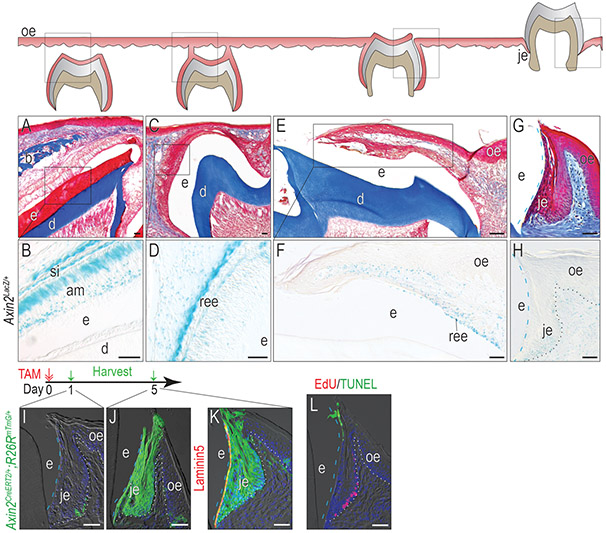
Figure 1. The JE originates from Wnt-responsive cells and maintains the Wnt-responsiveness.
(A) Masson’s trichrome staining showing the OE covering the unerupted maxillary first molar on P9. (B) In Axin2LacZ/+ mice, Xgal staining showing Xgal+ve Wnt-responsive cells in the JE on P9. (C) By P13, the enamel was completely formed, and the inner enamel epithelium reduced to a few layers of flat cuboidal cells called REE. At this stage, the REE started to fuse with OE. (D) Xgal staining showing Xgal+ve Wnt-responsive cells on P13. (E) On P14, the maxillary first molar penetrated to the oral cavity and the JE started to form. (F)Xgal staining showing Xgal+ve Wnt-responsive cells on P14. (G) Masson’s trichrome staining of the first maxillary molar in adult mice. (H) Xgal staining showing Wnt-responsive cells in adults. One dose of tamoxifen was given to adult Axin2CreERT2/+;R26RmTmG/+ mice and GFP+ve cells were analyzed (I) 1 day and (J) 5 days later. (K) Co-staining of 5-day chase GFP (green) with Laminin 5 (red) to show the attachment to the tooth surface. (L) TUNEL staining (green) showing apoptosis and EdU staining (red) showing cell proliferation in the JE.
Dashed blue lines indicate the shape of enamel. Dotted black and dotted white lines indicate the demarcation between the epithelium and the connective tissue. Abbreviations: e, enamel space; je, junctional epithelium; oe, oral epithelium; b, bone; d, dentin; ree, reduced enamel epithelium; am, ameloblasts. Blue dashed lines indicate the edge of the enamel and white dotted lines indicate the boundary between the epithelium and the connective tissue. Scale bars: 50μm.

