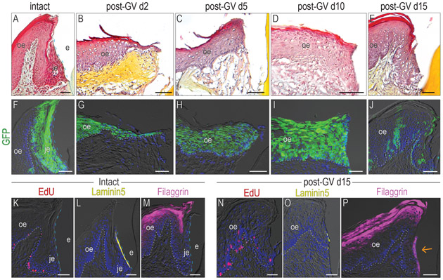
Figure 4. A functional JE cannot be solely regenerated from the OE.
Pentachrome staining was used to analyze the morphology with the samples from (A) intact group, post-GV day (B)2, (C) 5, (D) 10, and (E) 15. (F) One dose of tamoxifen was given to Axin2CreERT2/+;R26RmTmG/+ mice, GFP+ve cells were analyzed 5 days later. Lineage tracing showing the distribution of Wnt-responsive cells on post-GV day (G)2, (H) 5, (I) 10, (J) 15. In the intact samples, (K) EdU+ve cells, (L) Laminin 5 expression, and (M) Filaggrin expression pattern was examined. In the gingivectomy group, (N) EdU+ve cells, (O) Laminin 5 expression, and (P) Filaggrin expression pattern was examined on post-GV day 15. The orange arrow indicates the abnormal Filaggrin expression on the interface between the epithelium and tooth surface.
Dashed blue lines indicate the shape of enamel. Dotted white lines indicate the demarcation between the epithelium and the connective tissue. Abbreviations: GV, gingivectomy; je, junctional epithelium; oe, oral epithelium; e, enamel space. Scale bars: 50 μm.

