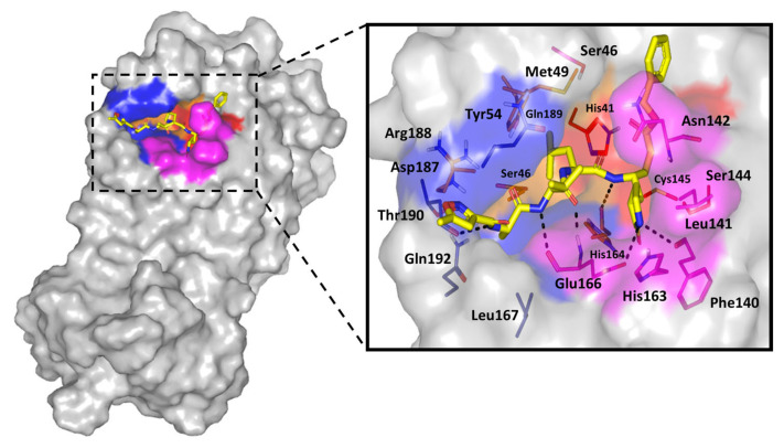
Figure 7.
Structure of crystallized SARS-CoV-2 3CLpro target complex (PDB code: 6lu7). Protein target (gray surface) is bound to the crystallized potent irreversible inhibitor (N3; yellow sticks), within the canonical substrate binding site. This shows the four important sub-pockets (S1’, S1, S2, and S3, as red, magenta, orange, and blue color, respectively). Zoomed stereoview of N3 (yellow sticks represent the ligand–protein hydrogen bonding as black dashed-lines. Residues (lines) located within 5 Å radius of bound ligands are colored in accordance with sub-pocket being labeled with sequence numbers.

