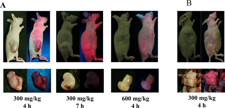
Fig 4. Fluorescent imaging of GIST xenograft models.
Images were captured using a high-resolution camera equipped with an optical filter. Each image was photographed under white light (left side) and LED illumination (right side). (A) Xenograft models of flank tumors. Strong fluorescence was observed in mice receiving 300 mg/kg 5-ALA for 4 h. (B) Xenograft models of peritoneal seeding tumors. Peritoneal dissemination could not be identified through skin via LED light, but strong fluorescence was observed in extracted tumors.

