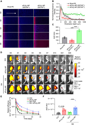
Fig. 3. Cartilage penetration and joint retention of sPLA2i-NPs.

(A) Representative confocal microscope images of cross sections of bovine cartilage explants incubated with free rhodamine dye or rhodamine-labeled sPLA2i-NPs in the presence or absence of cationic lipid DOTAP for 0, 2, 4, 6, and 8 days. Scale bar, 200 μm. (B) Semiquantitative analysis of the fluorescence intensity of the above different formulations over the entire explant sections after 8 days of incubation (n = 3). (C) Semiquantitative analysis of the AUC based on the fluorescence intensity profiles in (B) (n = 3). (D) Representative IVIS images of healthy and OA (OA was induced surgically 8 weeks before) mouse knee joints over 28 days after single intra-articular injection of free ICG or Cy7-labeled sPLA2i-NPs. (E) Semiquantitative analysis of time-course fluorescent radiant efficiency within healthy and OA mouse knee joints over 28 days (n = 5). (F) Semiquantitative analysis of the AUC based on the fluorescence intensity profiles in (E) (n = 5). Rhod-PE, free rhodamine dye; sPLA2i-NPs (DOTAP−), sPLA2i-NPs in the absence of DOTAP; sPLA2i-NPs (DOTAP+), sPLA2i-NPs in the presence of DOTAP. AU, arbitrary units. Statistical analysis was performed using one-way ANOVA with Turkey’s post hoc test. Data presented as means ± SEM. ***P < 0.001. Photo credit: Yulong Wei, University of Pennsylvania.
