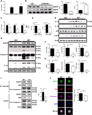
Fig. 1. GAT2 deficiency lowers the secretion of IL-1β and inhibits the activation of NF-κB signaling and inflammasome in proinflammatory macrophages.

(A) Relative mRNA expressions of SLC6A1, SLC6A13, SLC6A11, and SLC6A12 in wild-type (WT) resting macrophages (n = 4). Results represent two independent experiments. Data are shown as means ± SEM. (B) Protein expressions of GAT2 and GAT4 in unstimulated (US) or LPS/IFN-γ–stimulated WT macrophages (n = 3). Results represent two independent experiments. (C) Relative mRNA expressions of inducible nitric oxide synthase (iNOS), IL-1β, and IL-18 in WT and knockout (KO) proinflammatory macrophages (n = 3). Results represent two independent experiments. Data are shown as means ± SEM. (D) The secretion of IL-1β from LPS or plus IFN-γ–stimulated WT and KO macrophages (n = 4). Results represent three independent experiments. (E to H) Protein expressions of IL-1β, IL-18, NLRP3, caspase-1, p65, and p-p65 in WT and KO proinflammatory macrophages (n = 3 to 5). Results represent two independent experiments. (I) Immunoblot analysis of ASC immunoprecipitates in WT and KO proinflammatory macrophages, probed for NLRP3, pro–caspase-1, and ASC (n = 3). Results represent two independent experiments. IP, immunoprecipitation. (J) Confocal microscopy of WT and KO M1 macrophages, immunostained for NLRP3 (green), ASC (red), and Caspase-1 (purple) (n = 3). Scale bars, 10 μm. Data were analyzed by unpaired t test and represented as means ± SD unless indicated.*P < 0.05, **P < 0.0 , ***P < 0.001, and ****P < 0.0001. A.U., arbitrary unit; DAPI, 4′,6-diamidino-2-phenylindole.
