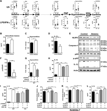
Fig. 4. GAT2 deficiency promotes betaine-hypoxanthine metabolism in proinflammatory macrophages.

(A) Schematic of the generation of hypoxanthine from betaine. Metabolites in this metabolic axis analyzed with metabolomics (n = 4 to 6), and the activities of PNP and XOD also measured (n = 4). (B to D) Intracellular betaine (B), hypoxanthine (C) and IL-1β secretion (D) in WT proinflammatory macrophages with betaine delivery (1 mM; n = 3). (E and F) Protein abundance of interested proteins in WT proinflammatory macrophages after betaine delivery (n = 3). (G and H) Intracellular hypoxanthine [(G) n = 6] and IL-1β secretion [(H) n = 3)] in LPS/IFN-γ–activated WT macrophages with betaine delivery combined BCX-1777 (10 μM) treatment. (I) IL-1β secretion from WT resting macrophages, LPS/IFN-γ–activated WT macrophages with PNP overexpression, and LPS/IFN-γ–activated KO macrophages with PNP silencing (n = 3). (J to L) The levels of extracellular (J) or intracellular (K) betaine and IL-1β secretion (L) in LPS/IFN-γ–activated RAW264.7 cells with GAT2 and/or GAT4 silencing in the presence of 50 μM betaine (n = 3). Results represent two (H and J to L), three (B to D), or four (G) independent experiments. Data were analyzed by unpaired t test (A, B, C, D, F, and G) or two-way ANOVA with Bonferroni correction (G to L) and represented as means ± SD. *P < 0.05, **P < 0.01, ***P < 0.001, and ****P < 0.0001.
