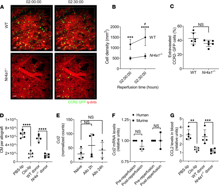
Figure 2. Depletion of donor nonclassical monocytes (NCM) suppresses the recruitment of recipient splenic classical monocytes (CM) to the allograft.
(A–C) Intravital 2-photon imaging between 2 and 2.5 hours after reperfusion. (A) Representative still images of WT and Nr4a1–/– donor grafts. Green, CCR2-GFP; red, Qdot655 blood vessels. (B and C) CCR2-GFP cell density (B) and percent of extravasated CCR2-GFP (C) calculated using NIH ImageJ software in WT and Nr4a1–/– mice grafts after transplantation. (D) Flow cytometry quantification of CM (live CD45+Ly6G–NK1.1–CD11b+SiglecF–CD24–Ly6Chi) recruited into the allograft after i.v. injection of PBS liposomes (PBS-lip) or clodronate liposomes (Clo-lip) in donor mice or using WT or Nr4a1–/– as donor lungs (n = 5). (E) Normalized counts per minute (CPM) of Ccl2 in sorted donor NCM isolated from allografts 2 and 24 hours after transplant. (F) Relative Ccl2 mRNA levels of human and mouse NCM isolated before and after reperfusion (n = 3). (G) CCL2 levels in blood from mice in D (n = 5). Graphs show means ± SD. The graph in B was analyzed by 2-way ANOVA followed by Sidak’s post hoc test. *WT versus Nr4a1–/–; #WT 2 hours versus WT 2.5 hours. The graph in E was analyzed by 1-way ANOVA followed by Tukey’s post hoc test. Graphs in C, D, F, and G were analyzed by unpaired Student’s t test. #P < 0.05, **P < 0.01; ***P < 0.001; ****P < 0.0001. Scale bar: 50 μm.

