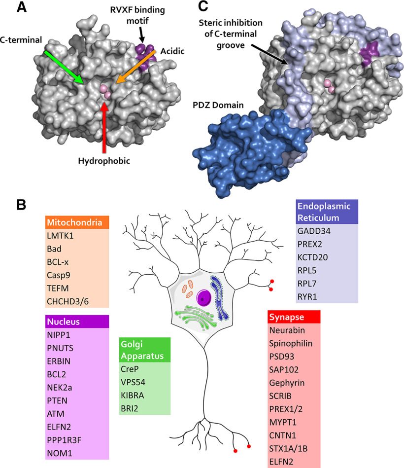
Figure 1.
PP1 active site and PP1 targeting. A, PP1α structure showing the active site (with two metal ions in light pink color) and the three grooves (colored arrows) radiating from the active site. B, Schematic showing examples of PP1 interacting proteins in various subcellular compartments in neurons. Localization data are based on the Human Protein Atlas (Uhlen et al., 2015; Thul et al., 2017) and postsynaptic density proteomics (Bayes et al., 2011). C, Example of PP1 targeting protein neurabin fragment binding to PP1α: in addition to using the RVxF binding mode (dark pink), neurabin (light purple) binds to the C-terminal groove of PP1, excluding a subset of PP1 substrates, such as phosphorylase α from the neurabin/PP1 holoenzyme (Ragusa et al., 2010). A, C, Structures were generated using PDB file 3hvq published by Ragusa et al. (2010).

