
Figure 2: Functional roles of CDK4 and CDK6 kinases.
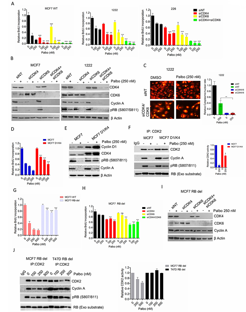
(A) BrdU incorporation in MCF7-WT, 1222 and 226 cell lines following the knockdowns of CDK4 and CDK6. Bars represent mean and SD (t-test) (B) Western blot analysis on MCF7 and 1222 cells following CDK4/6 knockdowns. (C) Representative images of 1222 cells stably expressing the CDK2 sensor following CDK4/6 concurrent knockdowns. Column graph indicates ratio of number of cells with cytoplasmic localization to number of cells with nuclear localization of the HDHB-mCHERYY. (D) BrdU incorporation in MCF7 and MCF7D1/K4 cells following 72 H exposure with palbociclib. Bars represent mean and SD (t-test) (E) Western blotting on MCF7 and MCF7D1/K4 cells following 48 H treatment with palbociclib (250 nM). (F) In vitro CDK2 kinase assays on MCF7 and MCF7D1/K4 cells treated with palbociclib (250 nM) up to 48 H. Representative blot images and mean and SEM are shown. (G) BrdU incorporation in MCF7-WT and MCF7-RB-del cells following 72 H exposure with palbociclib. Bars represent mean and SD (t-test). (H) BrdU incorporation in MCF7 RB depleted (MCF7 RB del) cell lines following the knockdowns of CDK4 and CDK6. Bars represent mean and SD (t-test). (I) Western blot analysis on MCF7 RB del following CDK4/6 knockdowns. (J) In vitro CDK2 kinase assays on MCF7 and T47D RB depleted cells treated with increasing concentrations of palbociclib up to 48 H. Representative blot images and mean and SEM are shown. Graphs represent 2 independent experiments with 3 replicates. (*p< 0.05, **p<0.01, ***p<0.001).