
Figure 3: Functional roles of P27 in PDAC and ER+ breast cancer models.
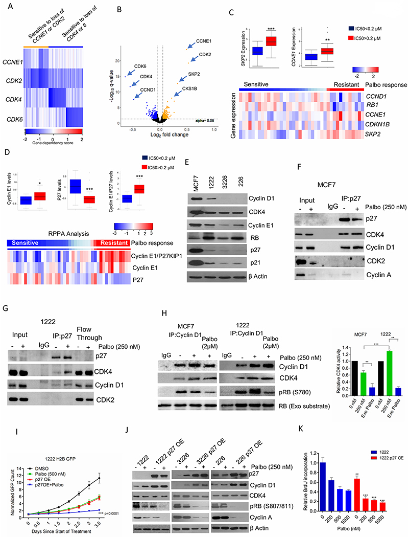
(A) Heat map represents the gene dependency score of different cancer cell lines that are sensitive to loss of CCNE1 (n=22), CDK2 (n=22) and to both CCNE1 and CDK2 (n=28) and their corresponding gene dependency scores to loss of CDK4 and CDK6 (Orange). Blue represents the top 50 cell lines that are only sensitive to loss of CDK4 and CDK6 independently and their corresponding gene dependency scores to loss of CCNE1 and/or CDK2. (B) Volcano plot indicates the differentially sensitive genes between the orange and blue groups, which are defined by a cut-off value +/− 0.05 for fold change and p-value 0.05 as determined by t-test. (C) Box plot representing the distribution of fold change of SKP2 and CCNE1 gene expression based on the IC50 values of palbociclib in different breast cancer cells. Heatmap indicates the distribution of fold change of the indicated genes based on the response to palbociclib. (D) Box plot and heat map representing the distribution of fold change of cyclin E1, P27 and cyclin E1/P27 based on the IC50 values of palbociclib in different breast cancer cells. (E) Western blot on the baseline expression of the indicated proteins from MCF7 and different PDAC cell lines. (F) Immunoprecipitation of P27 from MCF7 cells treated with palbociclib (250 nM) up to 48 H. Coimmunoprecipitated cyclin D1, CDK4, CDK2 and Cyclin A were determined by immunoblotting. (G) Immunoprecipitation of P27 from 1222 cells treated with palbociclib (250 nM) up to 48 H. Cyclin D1, CDK4 and CDK2 on the co-immunoprecipitated and flow through samples were determined by immunoblotting. (H) In vitro CDK4 kinase assay associated with cyclin D1 on MCF7 and 1222 cells treated with palbociclib (250 nM) up to 48 H. Exogenous palbociclib (2 μM) was added to the kinase reaction mix. Kinase activity was evaluated based on the phosphorylation of an exogenous RB fragment as substrate at S780. Representative blot images and mean and SD are shown. (I) Growth rate of 1222-WT and 1222 P27 OE cells treated with palbociclib (500 nM). Bars represent mean and SD (2-way ANOVA). (J) Immunoblot analysis on 1222, 226 and 3226 cell lines and their respective P27 OE cells in the presence of palbociclib (250 nM) up to 48 H. (K) BrdU incorporation of 1222 and 1222 P27 OE cells following 72 H exposure in the presence of palbociclib. Bars represent mean and SD (t-test). (*p< 0.05, **p<0.01, ***p<0.001).