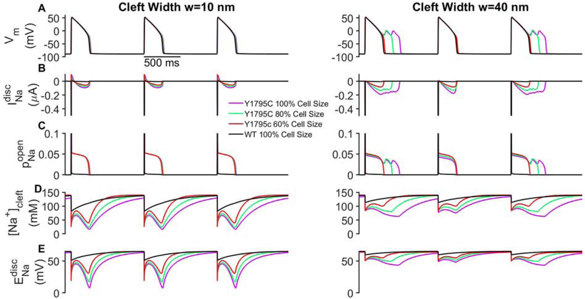
Figure 2:

Early after-depolarizations (EADs) depend on cleft width and cell size. (A) Transmembrane voltage (Vm), (B) Na+ current at the ID , (C) Na+ channel open probability at the ID , (D) cleft Na+ concentration ([Na+]cleft), and (D) the Na+ reversal potential at the ID are shown in mutant tissue for narrow (left) and wide (right) intercellular cleft widths (w). Traces for wild-type (WT) are shown for 100% cell size for comparison (black lines). For clarity, traces are shown for cell 25 out of 50 in the one-dimensional chain of cells. Parameters: BCL = 1000 ms, IDNa = 50%, ρNa = 100%, fgap = 100%.
