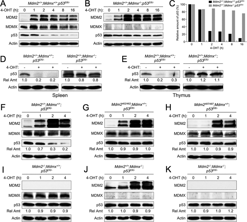
Figure 1. Deletion of MDMX in mice causes p53 accumulation.
Early passages of (A) Mdm2+/+;Mdmx+/+;p53ER/- and (B) Mdm2+/+;Mdmx−/−;p53ER/- mouse embryonic stem (MEF) cells were treated with 4-hydroxytamoxifen (4-OHT) for the indicated times. The levels of MDM2, MDMX, p53 and actin were analyzed by western blot. (C) The amounts of p53 remained at each time point in A-B were quantified by densitometry, normalized to actin, and plotted. (D) Spleen and (E) thymus tissues were isolated from 8 weeks old Mdm2+/+;Mdmx+/+;p53ER/- and Mdm2+/+;Mdmx−/−;p53ER/- mice after treatment with 4-OHT for 6 hours. Tissue lysates were analyzed by western blot. Relative amounts of p53 were indicated. (F-K) MEFs of varies MDM2 and MDMX statuses were treated with 4-OHT for the indicated times and cell lysates were analyzed by western blot. Relative amounts of p53 were indicated.

