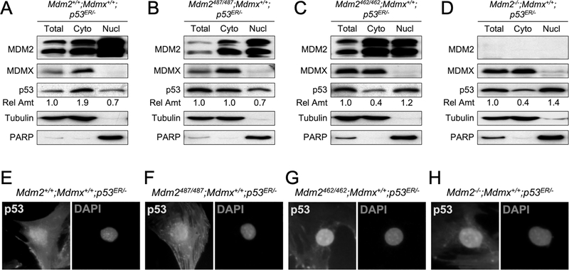
Figure 4. MDM2-MDMX heterodimerization facilitates p53 cytoplasmic localization.
Cell lysates were isolated from (A) Mdm2+/+;Mdmx+/+;p53ER/-, (B) Mdm2487/487;Mdmx+/+;p53ER/-, (C) Mdm2462/462;Mdmx+/+;p53ER/-, and (D) Mdm2+/+;Mdmx−/−;p53ER/- MEFs. The lysates were either untreated (Total) or separated into cytoplasmic (Cyto) and nuclear (Nucl) fractions and analyzed for MDM2, MDMX, and p53 by western blot. Tubulin and PARP were used as controls for cytoplasmic fraction and nuclear fraction, respectively. The relative amounts of p53 in each fraction were shown. (E-H) MEFs of indicated genotypes were stained with p53 antibody and counter-stained with DAPI, and cell images were taken by microscope. Representative images were shown.

