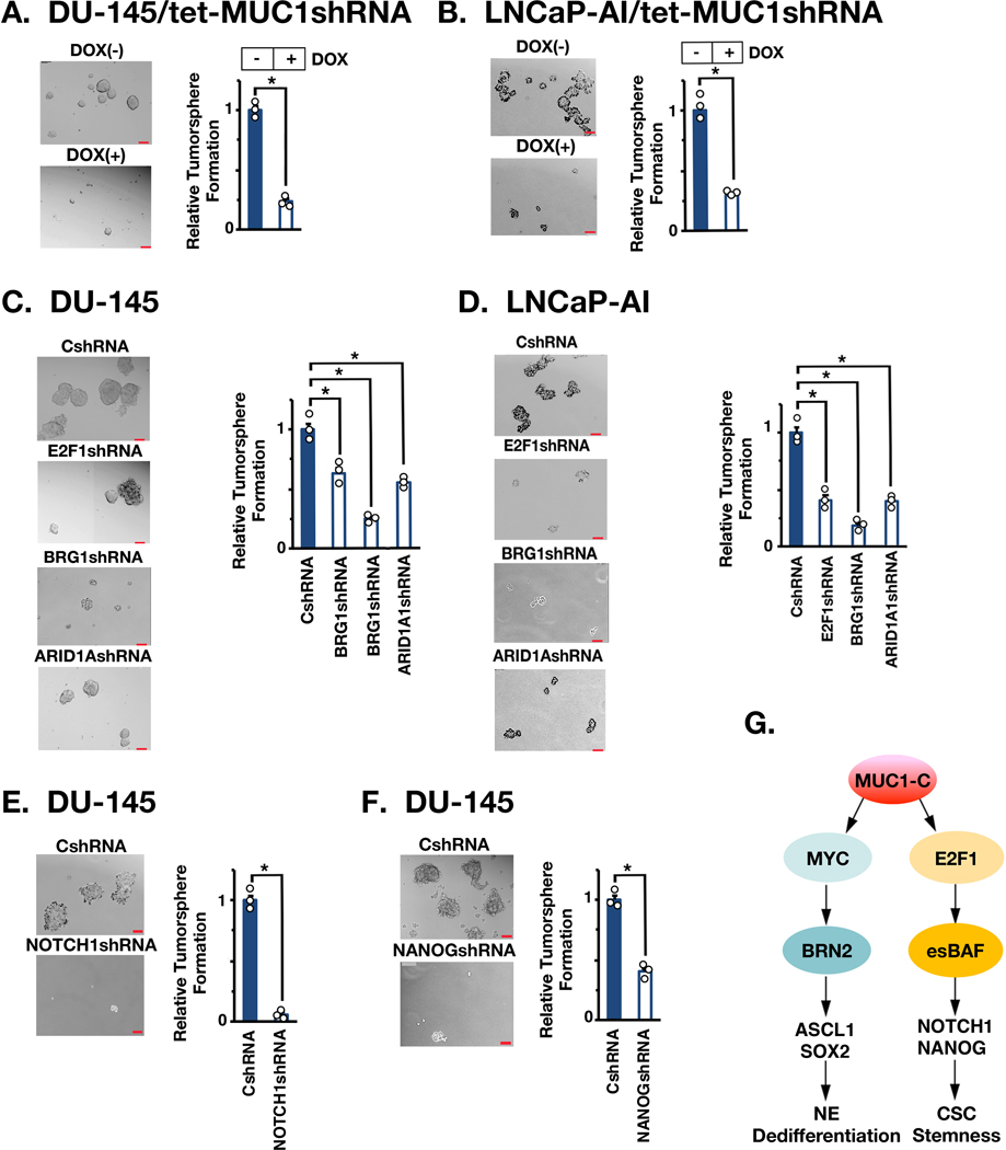
Figure 7. MUC1-C→E2F→BAF→NOTCH1→NANOG pathway is necessary for self-renewal.
A and B. DU-145/tet-MUC1shRNA (A) and LNCaP-AI/tet-MUC1shRNA (B) cells treated with vehicle or DOX for 10 days were assayed for tumorsphere formation (left). Scale bar: 100 mm. The results (mean±SD of 3 biologic replicates) are expressed as relative tumorsphere number per field compared to the CshRNA control (right). C. The indicated DU-145 cells were assayed for tumorsphere formation (left). The results (mean±SD of 3 biologic replicates) are expressed as relative tumorsphere number per field compared to the CshRNA control (right). D. The indicated LNCaP-AI cells were assayed for tumorsphere formation (left). The results (mean±SD of 3 biologic replicates) are expressed as relative tumorsphere number per field compared to the CshRNA control (right). E and F. DU-145 cells expressing a CshRNA, NOTCH1shRNA (E) or NANOGshRNA (F) were assayed for tumorsphere formation (left). The results (mean±SD of 3 biologic replicates) are expressed as relative tumorsphere number per field compared to the CshRNA control (right). G. Proposed model for involvement of MUC1-C in integrating MYC and E2F1 signaling pathways in NEPC progression. MUC1-C binds directly to MYC and drives activation of BRN2, the Yamanaka OSK pluripotency factors and NE dedifferentiation (10). The present results demonstrate that MUC1-C binds directly to E2F1 and activates the esBAF complex (BRG1, ARID1A, BAF60a, BAF155 and BAF170). The MUC1-C→E2F1→esBAF pathway drives the induction of NOTCH1, NANOG and CSC self-renewal.

