
Figure 1-. Murine model of resection including dexamethasone treatment extends survival and reduces edema.
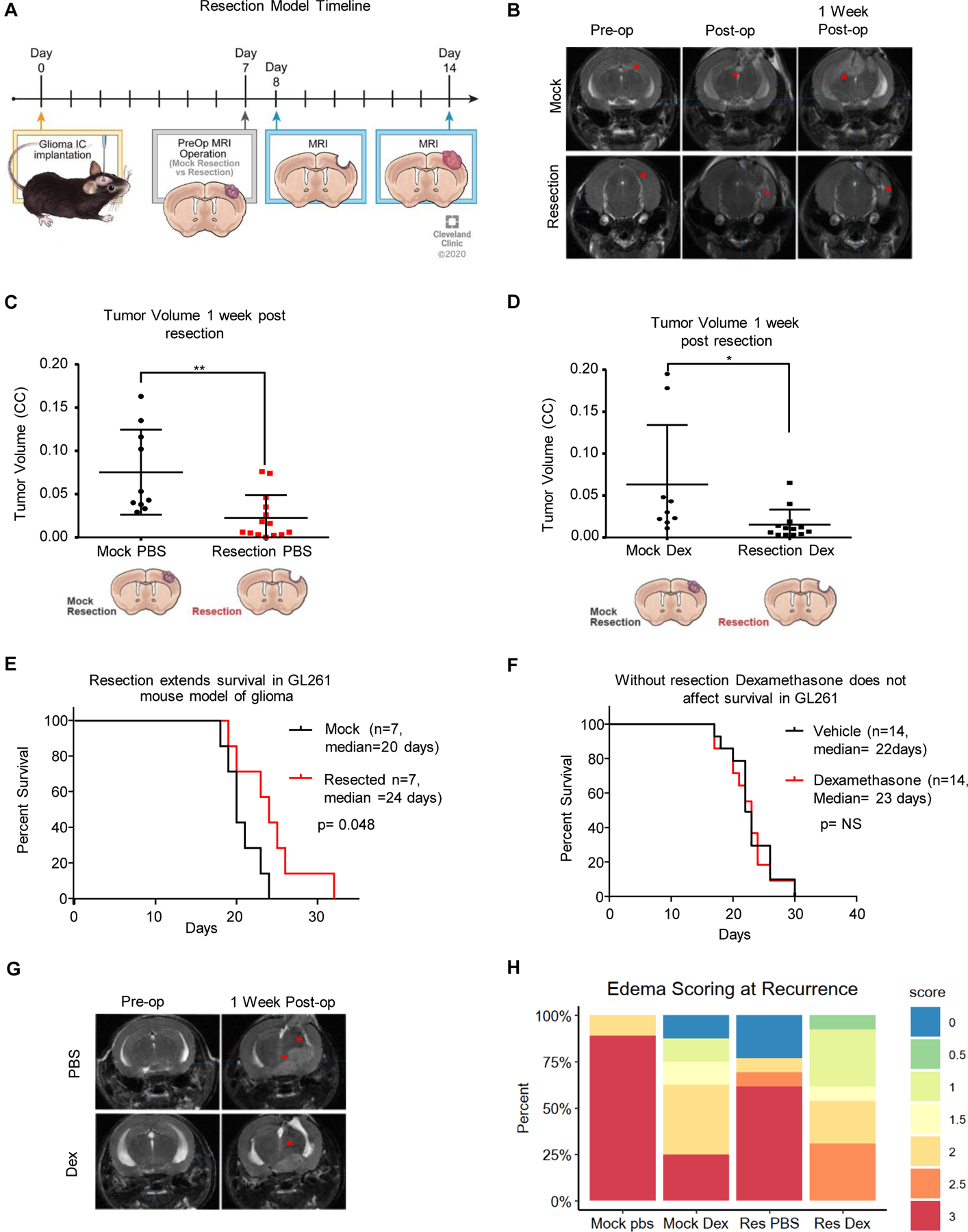
To replicate standard-of-care and dexamethasone treatment, a mouse model of resection was initiated as outlined in the diagram, with intracranial implantation of tumor, followed by MRI prior to resection and after resection, and then endpoint MRI along with flow cytometry or survival depending on the experiment (A). Representative MRI images of tumors from the mock resection and resection cohorts pre-, post- and 1 week post-resection (B). Tumor volume was assessed using the BrainLab software suite and graphed as tumor volume (C, D). A survival study comparing n=7 mice with mock resection and n=7 mice with resection was performed showing a median survival of 20 days for mock resection vs 24 days for resection, with log-rank p-value shown (E). Vehicle- vs dexamethasone (one week at 4 µg daily)-treated GL261-bearing mice with no surgical resection were also compared and showed no survival difference due to dexamethasone (F). Representative MRI images of dexamethasone-treated mice and PBS-treated mice pre-op and 1 week post-op (G). Edema scoring is graphed as a percentage with n=7 mice per group (H). Student’s two-tailed t-test was performed for comparisons in panels A, D, E; *p<0.05, **p<0.01, ***p<0.001. Survival curve analysis was performed in GraphPad Prism using log-rank tests (also known as Mantel-Cox tests) for p values.