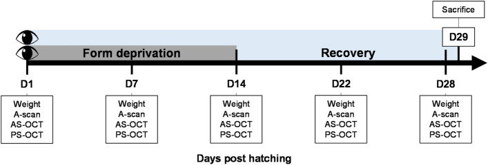
Figure 2.
Experimental protocol. Thirty-six, 1-day old chicks were randomly assigned to two groups of 18 animals each and raised under SW or BEW light emitting diode (LED) lighting for 29 days. Animals were randomly monocularly fitted with a diffuser on D1. Diffusers were removed on D14 and FDEP eyes recovered until D29. Ophthalmic examinations were performed on D1, D7, D14, D22 and D28. On D29, animals were sacrificed and eyes were enucleated. The retinas and vitreous were harvested for metabolomic analysis. Ten eyes were dissected for histological evaluation of the retina, choroid and sclera. AS-OCT anterior segment optical coherence tomography, PS-OCT posterior segment optical coherence tomography.

