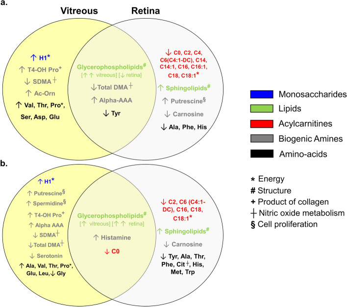
Figure 8.
Venn diagrams summarizing metabolomic changes in the vitreous and retina of recovering FDEP (a) and control (b) eyes in animals exposed to BEW light compared to SW light. Compared to eyes reared under SW light, the metabolomic profiles within the FDEP and control eyes reared under BEW, involved an increase in vitreal H1, a reduction in retinal acylcarnitines, as well as increases in glycerophospholipids and sphingolipids in the vitreous and retina, respectively. Recovering FDEP eyes and control eyes exposed to BEW light also exhibited changes in biogenic amines (e.g., total and symmetric dimethylarginine (total DMA and SDMA), putrescine, spermidine, alpha aminodipic acid (alpha-AAA) and serotonin) and amino acids (e.g., alanine, valine, threonine, histidine and tyrosine) in the vitreous and retina. Alpha-AAA alpha-aminodipic acid, Ala alanine, Asp aspartate, C0 carnitine, C2 acetylcarnitine, C4 butyrylcarnitine, C6 hexanoylcarnitine, C6 (C4:1-DC) hexanoylcarnitine (fumarylcarnitine), C14 tetradecanoylcarnitine, C14:1 tetradecenoylcarnitine, C16 hexadecanoylcarnitine, C18 octadecanoylcarnitine, C18-1 octadecenoylcarnitine, Cit citrulline, Glu glutamate, H1 sum of hexoses (including glucose), His histidine, Iso isoleucine, Lys lysine, Met methionine, Phe phenylalanine, Pro proline, SDMA symmetric dimethylarginine, t4-OH Pro trans-4-hydroxyproline, Thr threonine, Total DMA total dimethylarginine, Trp tryptophan, Tyr tyrosine, Val valine.

