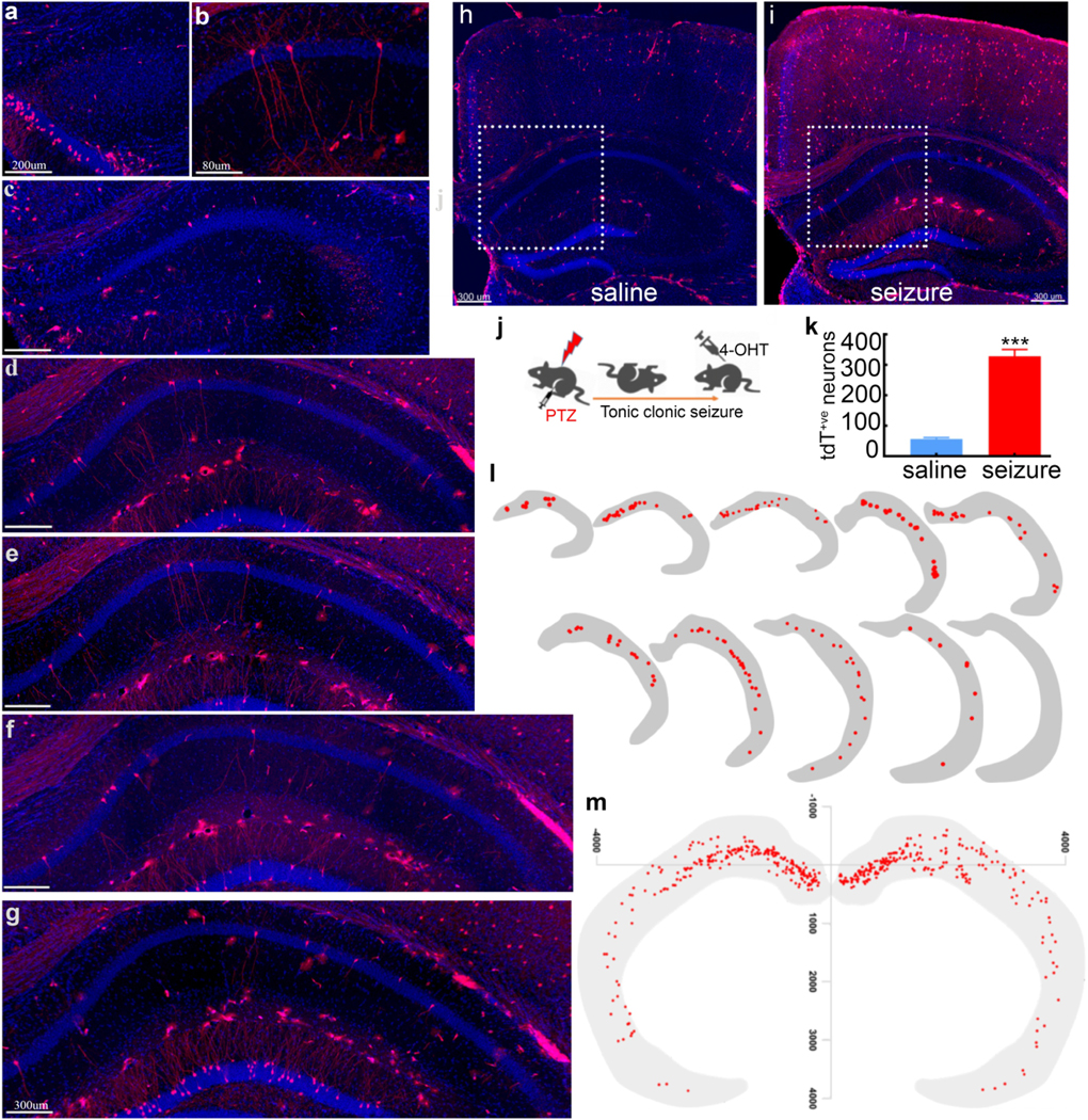
Fig. 3. Seizure-tagged CA1 neuronal ensemble map is similar to memory map:

a-i) Representative images show tagged CA1 pyramidal neurons (red) activated following a seizure across the anterior-posterior axis, with DAPI (blue) counterstaining. Anterior dorsal CA1 had more tagged neurons than the ventral part. h) There were fewer tdT+ve neurons in representative images of the hippocampus from saline-injected mice, compared to seizure treated mice. There were one to two tagged CA1 pyramidal neurons (dotted square) per 40μm hippocampal slice. (i), with 5–7 tagged pyramidal neurons in distal dCA1 (dotted square). j) The experimental design for tagging neurons by a seizure. k) Seizure tagged more CA1 pyramidal neurons than saline injection (n=6, ***p<0.001). l) Unilateral location maps (10) representing 50 (40μm) hippocampal slices show the distribution of seizure-tagged pyramidal CA1 neurons rostrocaudal axis. m) Combined location map of seizure-tagged CA1 ensemble showing their distribution across the rostrocaudal and medial-lateral axis.
