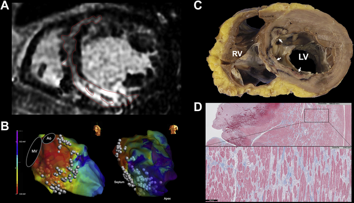
Figure 7.
Pathologic analysis of septal scar and ablation lesion. A: FS patient with C-shaped pattern underwent heart transplantation 4 days after a fourth unsuccessful ablation procedure. B: Ablation along the entire septum and LV inferior wall with low unipolar voltages shown on electroanatomic mapping. C: Explanted heart showed incomplete penetration of multiple ablation lesions into the midseptum (white arrows). D: Trichrome stain showing extensive midseptal fibrosis with interspersed viable myocardium. Ao = aorta; MV = mitral valve; other abbreviation as in Figure 1.

