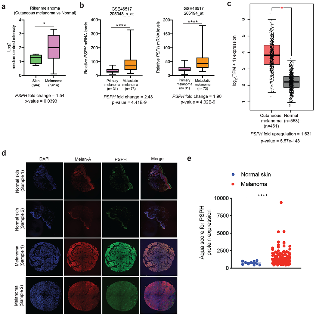
Fig. 1. PSPH is overexpressed in melanoma.

a. The indicated melanoma datasets were analyzed for PSPH mRNA expression. The relative PSPH mRNA expression in patient-derived melanoma samples was compared with normal skin. b. Comparison of PSPH expression in patient-derived melanoma samples from subjects with metastatic melanoma and those with primary melanoma. c. Comparison of PSPH mRNA expression in TCGA melanoma samples with GTEx and TCGA normal samples combined. d. Quantitative immunofluorescence analysis of Tissue Microarray (TMA) with melanoma and normal skin samples. Representative AQUA immunofluorescence images of the indicated melanoma cell type and normal skin samples. Samples are stained for DAPI, Melan-A, and PSPH as indicated. e. The average AQUA scores for melanoma and normal skin samples are plotted and presented as the mean ± standard error of the mean (SEM). * and **** represent P < 0.05 and P < 0.0001, respectively.
