
Fig. 2. PSPH is necessary for melanoma tumor growth and metastasis.
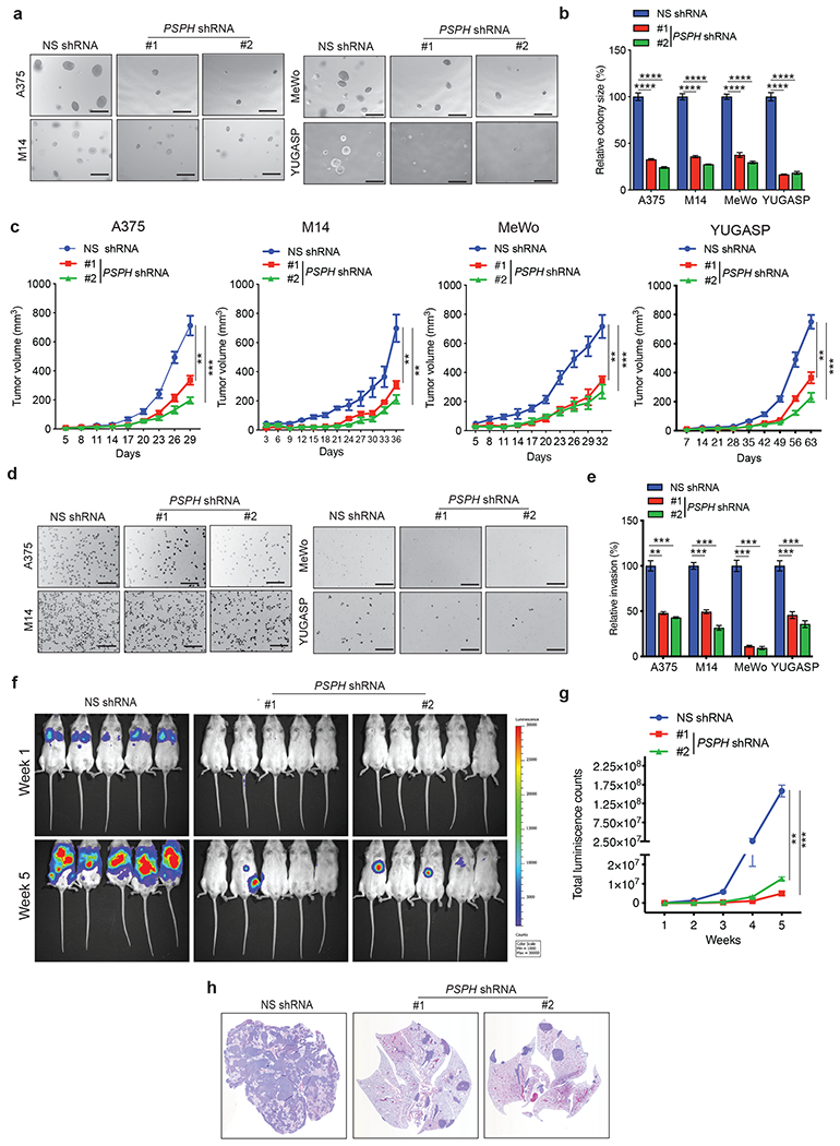
a. Anchorage-independent growth was measured using the soft-agar assay in indicated cell lines expressing either PSPH short hairpin RNA (shRNA) or a nonspecific (NS) control shRNA. Representative images of soft-agar colonies from the indicated melanoma cell lines are shown. Scale bar, 500 μm. b. Plot showing relative colony sizes from the soft-agar assay presented in panel A. c. Indicated melanoma cell lines expressing either PSPH shRNA or NS shRNA were subcutaneously injected into the flanks of athymic nude mice (n = 3). Average tumor volumes at the indicated time points are shown. d. Matrigel invasion assays with the indicated melanoma cell lines expressing PSPH shRNA or NS shRNA; representative images are shown. Scale bar, 200 μm. e. Relative invasion (%) from Matrigel assays shown in panel D. f. A375-MA2-F-Luc cells expressing PSPH shRNA or NS shRNA were administered to NSG mice (n = 5) via tail vein injection. Bioluminescence images of mice from the indicated groups at weeks 1 and 5 are shown. g. Quantitation of bioluminescence in the mice at the indicated time points. h. Representative images of hematoxylin-and-eosin (H&E)–stained lung sections showing the histology of lungs with tumor metastasis. Data are presented as the mean ± SEM; **, ***, and **** represent P < 0.01, P < 0.001 and P < 0.0001, respectively.