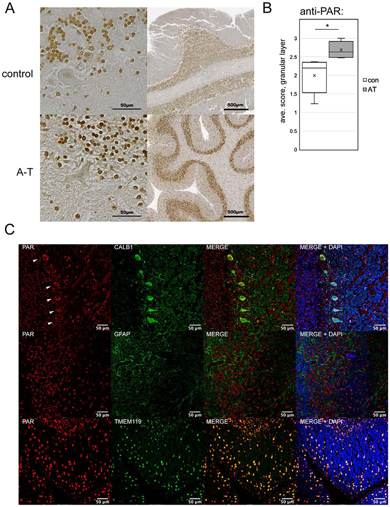
Figure 7. Elevated PAR is present in A-T patient cerebellum tissue.
(A) Examples of cerebellum tissue (formalin-fixed) from an A-T patient (#6) and control, analyzed by immunohistochemistry with an antibody directed against poly-ADP-ribose, HRP-conjugated secondary antibody, and 3,3'Diaminobenzidine with methyl green counterstain at two resolutions as indicated. (B) Subjective scoring of the level of PAR staining of cells in the granular layer of 5 A-T patient cerebellum samples (5, 6, 12, 18, 21) compared to age-matched controls was performed using a 0 to 3 scale of blinded samples by 3 individuals. Error bars indicate standard deviation. *, **, ***, and **** indicate p<0.05, 0.005, and 0.0005 by Student two-tailed t-test; NS = not significant. (C) Examples of A-T patient formalin-fixed cerebellum tissues stained with antibodies directed against PAR, CALB1 (Purkinje cells), GFAP (astrocytes), and TMEM119 (microglia) as indicated from A-T patients #5, 12, and 1, respectively, using fluorescence-labeled secondary antibodies and DAPI. White arrows indicate Purkinje cells in the CALB1/PAR-stained images.

