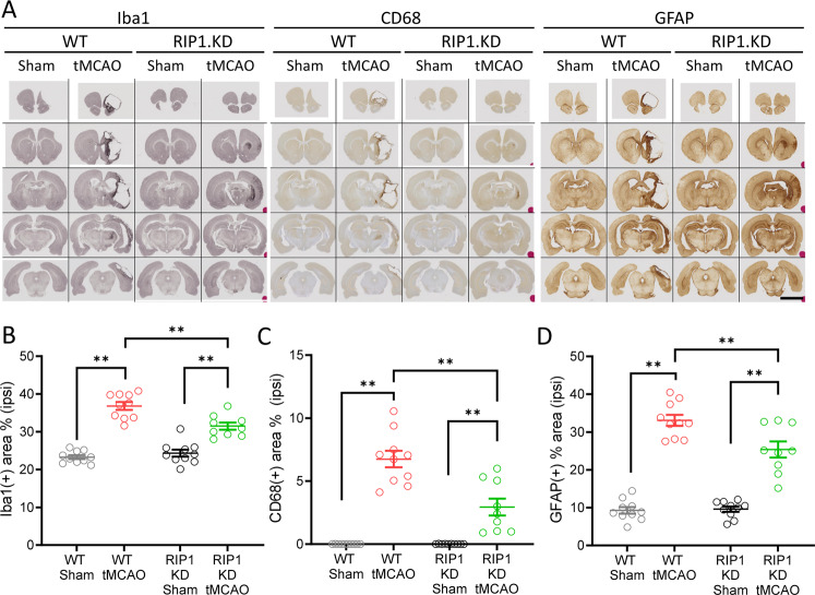
Fig. 4. Genetic inactivation of RIP1 kinase activity reduces inflammation following ischemic brain injury.
A Immunohistochemistry for Iba1, CD68, and GFAP in WT and RIP1 KD rats at 30-days post-tMCAO. Representative images across the brain are shown. Scale bar, 5 mm. B–D Quantification of percent tissue area positive for high-intensity Iba1 (B), CD68 (C) and GFAP (D) in the ipsilateral side, from (A). n = 8–10 rats/group. **p < 0.01 by Two-way ANOVA (Holm-Sidak). Data are represented as mean ± S.E.M.

