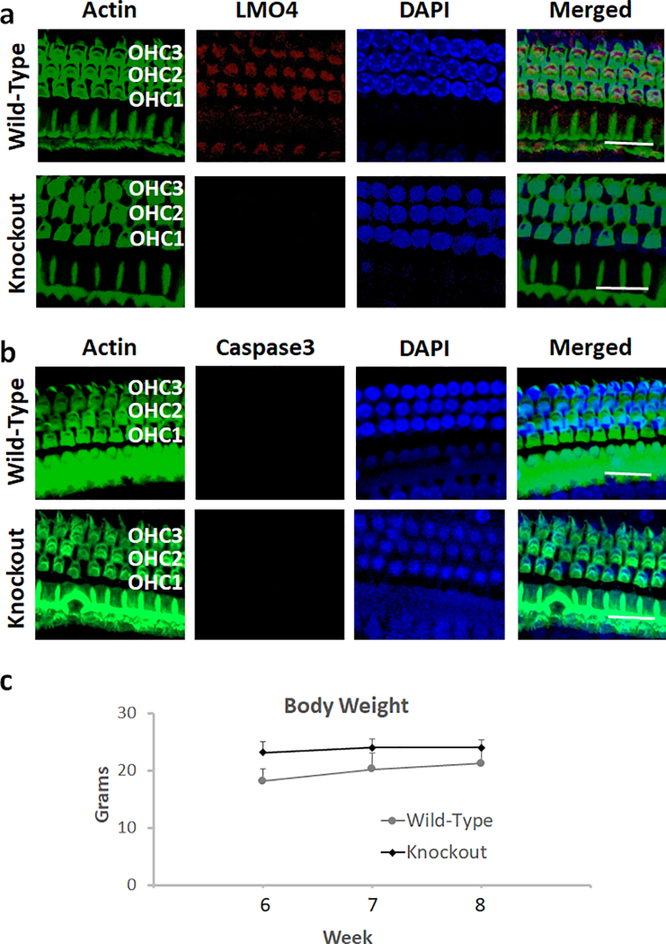
Fig. 2.
Deletion of LMO4 and apoptosis in the hair cells of knockout mice. a) Immunolocalization with anti-LMO4 indicated the expression of LMO4 (red stain) in the outer hair cells of wild-type mice. However, the expression of LMO4 was not detected in the outer hair cells of knockout mice. Green indicates staining of actin with phalloidin while blue indicates staining of the nucleus with DAPI. Images are representative of three replicates. Scale bar = 20 μm. b) Immunolocalization with anti-Caspase 3 indicated that the expression of activated caspase 3 (red stain) in the outer hair cells of wild-type and knockout mice was very low. Green indicates staining of actin with phalloidin while blue indicates staining of the nucleus with DAPI. Images are representative of three replicates. Scale bar = 20 μm. c) Measurement of the body weight at 6, 7 and 8 weeks indicated that the weight gain of knockout mice was similar to that of the wild-type. The results are expressed as mean ± standard deviation, n = 6.

