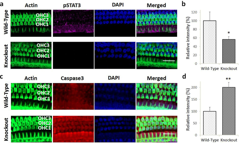
Fig. 5.
Effect of Lmo4 deletion on cisplatin-induced inactivation of STAT3 and apoptosis in the hair cells. a) Immunolocalization with anti-pSTAT3 indicated that cisplatin-induced decrease in the expression levels of phosphorylated STAT3 (magenta stain) in the knockout mice was greater than that observed in the wild-type littermates. Green indicates staining of actin with phalloidin while blue indicates staining of the nucleus with DAPI. Images are representative of five replicates. Scale bar = 20 μm. b) Quantification of the immunostaining indicated that the cisplatin-induced decrease in the expression of pSTAT3 in the knockouts was significantly lower than that of wild-type littermates. The results are expressed as mean ± standard error mean, n = 5 (*p = 0.0395). c) Immunolocalization with anti-Caspase 3 indicated that cisplatin treatment induced the expression of activated caspase 3 (red stain) in the outer hair cells of wild-type and knockout mice. However, cisplatin-induced increase in the expression levels of activated caspase 3 in the knockout mice was much higher than that observed in the wild-type littermates. Green indicates staining of actin with phalloidin while blue indicates staining of the nucleus with DAPI. Images are representative of five replicates. Scale bar = 20 μm. d) Quantification of the immunostaining indicated that the cisplatin-induced increase in the expression of activated caspase 3 in the knockouts was significantly higher than that of wild-type littermates. The results are expressed as mean ± standard error mean, n = 5 (**p = 0.0026).

