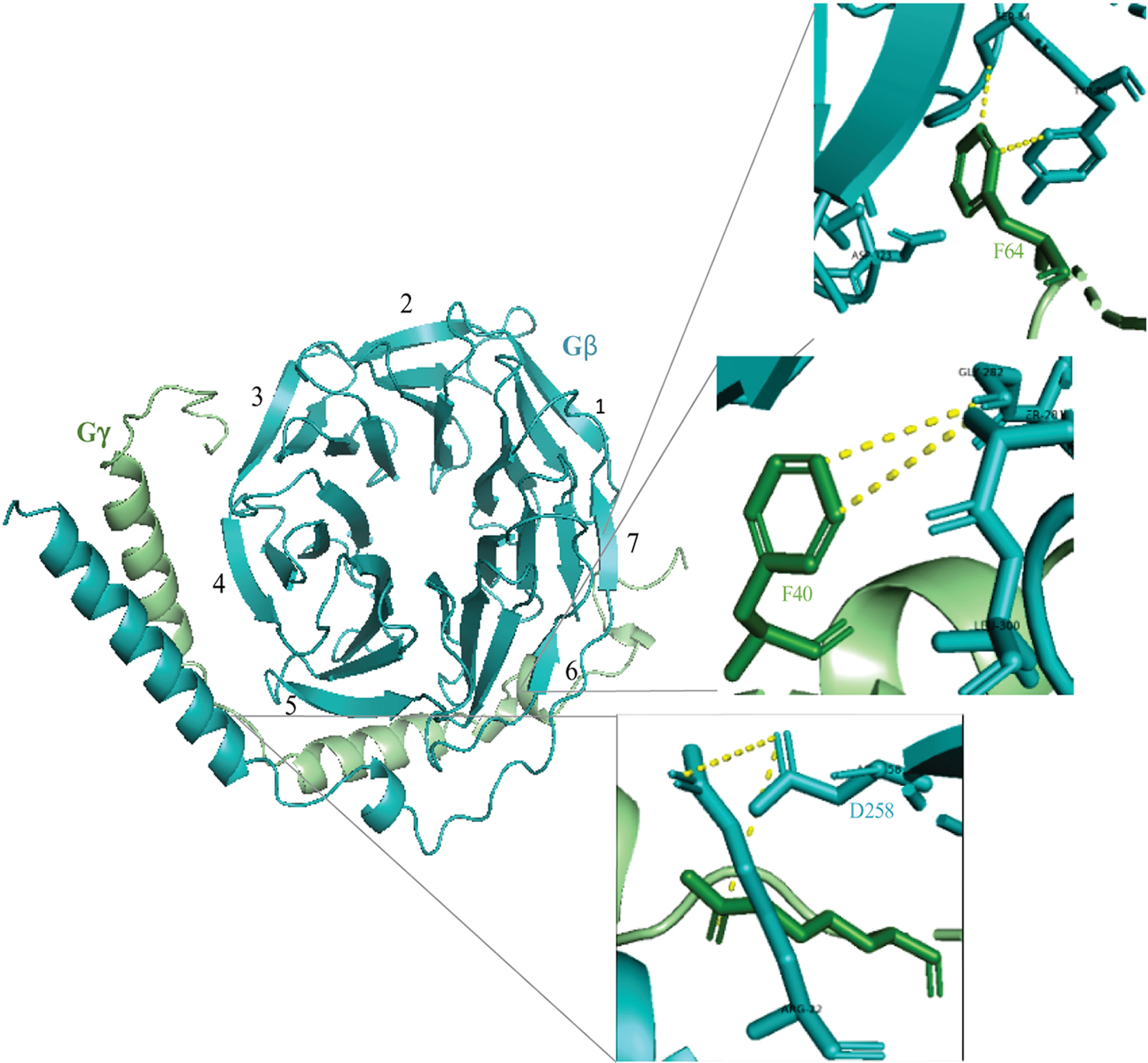
Figure 2. Assembly of Gβ1γ1 dimer crystal structure adapted from the PDB ID 1TBG.

Gβ and Gγ subunits are shown in cyan, blue and green colors respectively. Black numerals indicate seven blades of the Gβ propeller. A ribbon representation of the dimer indicates some of the interaction sites between the subunits. The magnified diagram shows interaction of Gβ-Asp258 with residues on both the subunits. Hydrophobic interactions between Gγ residues including Phe40 and Phe64 with Gβ are also labeled.
