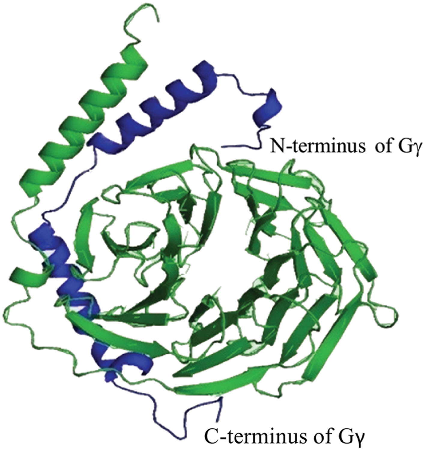
Figure 8. Molecular model structure of Gβγ dimer of Gβ1 and Gγ1, using coordinates obtained from PDB file 1TBG.

Gβ1 and Gγ1 are shown in green and blue respectively. The N-terminal helix of Gγ1 interacts with the N-terminal helix of Gβ1 to form a coiled-coiled structure. The C-terminal helix of Gγ1 shows interactions with the β-propeller domain of Gβ1.
