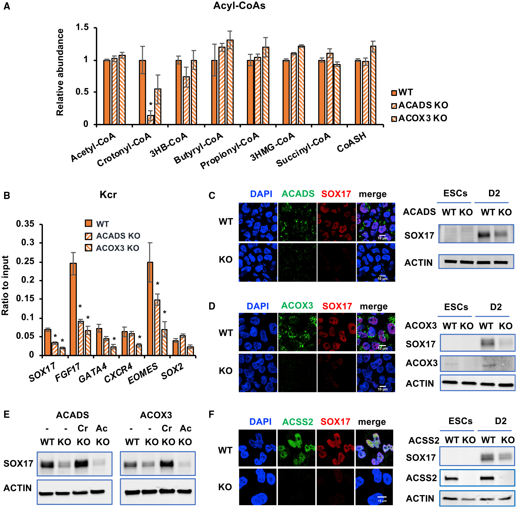
Figure 6. Crotonyl-CoA-producing enzymes modulate histone crotonylation and regulate endoderm differentiation in vitro.

(A) KO of ACADS or ACOX3 reduces crotonyl-CoA levels. The levels of crotonyl-CoA in the indicated mel1-differentiated D3 endodermal cells were measured as described in Method details (n = 3 biological replicates/group, *p < 0.05, values are expressed as mean ± SEM).
(B) Knocking out ACADS or ACOX3 in mel1 hESCs reduces deposition of Kcr at the TSSs of endodermal genes. TSS-associated Kcr levels were analyzed by ChIP-qPCR in the indicated cells, and relative enrichment was calculated against the input signal (n = 3 independent experiments, *p < 0.05, values are expressed as mean ± SEM).
(C) Knocking out ACADS in mel1 hESCs impairs endodermal gene expression. Left: the expression of ACADS and SOX17 was analyzed in WT and ACADS KO D2 endodermal cells by immunofluorescence staining. Right: the expression of SOX17 was analyzed in hESCs and the indicated D2 endodermal cells by immunoblotting. Scale bars, 10 μm.
(D) Knocking out ACOX3 in mel1 hESCs impairs endodermal gene expression. WT and ACOX3 KO D2 endodermal cells were analyzed as in (C). Scale bars, 10 μm
(E) Crotonate, but not acetate, rescues the defective endoderm differentiation induced by ACADS or ACOX3 deficiency. WT and the indicated KO mel1 hESCs were induced into endoderm differentiation in the absence or presence of 5 mM crotonate (Cr) or acetate (Ac) for 2 days.
(F) Deletion of ACSS2 in mel1 hESCs impairs endodermal gene expression. WT and ACSS2 KO D2 endodermal cells were analyzed as in (C). Scale bar, 10 μm.
See also Figure S6.
