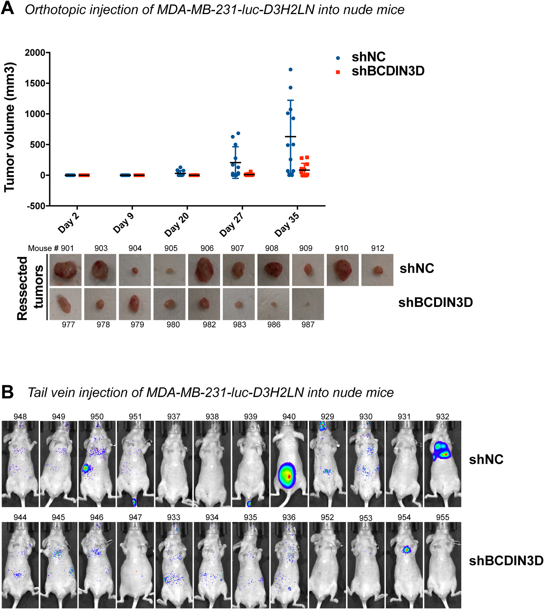
Figure 1. BCDIN3D depletion reduces tumor formation in vivo.

a Foxn1nu/nu mice were orthotopically injected into the #4 mammary fat pad with MDA-MB-231-luc-D3H2LN shNC or shBCDIN3D cells. Tumors were palpated and measured with a caliper over a period of five weeks. The graph shows the tumor volume (mm³) calculated by the formula: volume = (smaller dimension² × larger dimension)/2 as a function of time (scatter plot with mean ± SD, n=10 for shNC, and n=8 for shBCDIN3D, please note that the mice who did not develop tumors are shown on the graph with a dot at 0, but were not taken into account for the calculation of the average size of tumors). The pictures show the resected tumors at day 35 post-injection.
b Shown are IVIS images of mice injected with luciferin intraperitoneally 8 weeks post-tail vein injection of MDA-MB-231-luc-D3H2LN shNC or shBCDIN3D cells. 3 control mice show tumors on the bone [spine (#940), rib cage (#950) and jaw (#929)], 1 control mouse (#932) shows a large tumor in the lungs and 1 mouse injected with shBCDIN3D cells (#954) shows a smaller tumor in the lungs.
