FIGURE 2.
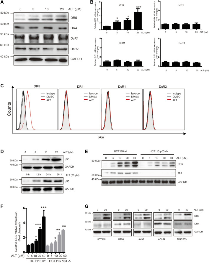
Alternol induces expression of DR5. (A) Caki-1 cells were treated with various doses of alternol (ALT) for 24 h. Total cellular extracts were then prepared and analyzed for DR4, DR5, DcR1, and DcR2 by western blot. (B) Caki-1 cells were treated with various doses of alternol (ALT) for 24 h. DR5, DR4, DcR1, and DcR2 mRNA levels were analyzed by qRT-PCR. (C) Cellular surface levels of DR5, DR4, DcR1, and DcR2 were analyzed by flow cytometry staining using PE-conjugated specific antibodies for each receptor. IgG isotype controls (gray histogram), DMSO (gray line), and alternol (red line) are presented. (D) Caki-1 cells were treated with various doses of alternol for 24 h or treated with 20 μM alternol for different times. Then, cellular lysates were analyzed with indicated antibodies. HCT116 wt and HCT116 p53−/− cells were treated with various doses of alternol for 24 h, and DR5 protein and mRNA levels were analyzed by western blot (E) and qRT-PCR (F), respectively. (G) Various types of carcinoma cells were treated with 20 μM alternol for 24 h, after which whole-cell extracts were analyzed by western blot assay. The same blots were stripped and re-incubated with actin antibody to confirm equal protein loading. Data are the mean ± SD of three independent experiments. *p < 0.05; **p < 0.01; ***p < 0.001, compared with 0 μM ALT.

