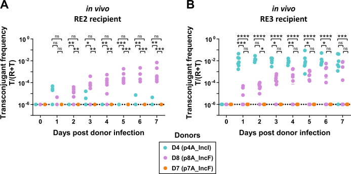
Fig. 4. ESBL-plasmids can spread in the gut in the absence of antibiotic selection.
We measured the spread of three plasmids as final transconjugant frequency in two distinct recipient populations, A RE2 and B RE3, and enumerated transconjugants in faeces by selective plating. Dotted lines indicate the detection limit for selective plating. Circles represent independent replicates (n = 7 for RE2 conjugations; n = 7 for D4-RE3; n = 10 for D8-RE3 and D7-RE3), lines show the median and different donor-plasmid pairs are indicated in colour. Kruskal–Wallis test p > 0.05 (ns), p < 0.05 (*), p < 0.01 (**), p < 0.001 (***), p < 0.0001 (****). Total population densities can be found in Supplementary Fig. S14.

