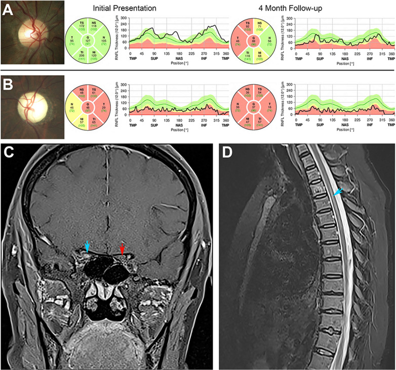
Fig. 2. Clinical presentation of a 49-year-old female with sequential bilateral NMOSD-ON.
The patient presented with acute vision loss to hand motions in the right eye, 3 months after having lost vision to no-light perception in the left eye. Visual acuity improved to 20/25 in the right eye following intravenous methylprednisolone and plasma exchange therapy. She was subsequently treated with intravenous rituximab infusions every 6 months. A The right optic disc (left panel) appeared normal on presentation, with minimal thickening of the peripapillary retinal nerve fibre layer (pRNFL) on optical coherence tomography (OCT; middle panel). Four months later, substantial pRNFL thinning had developed, particularly in the temporal quadrant (right panel). B The left optic disc exhibited considerable pallor, and pRNFL was severely thinned on initial presentation, consistent with profound optic atrophy. Some additional interval thinning was apparent 4 months later. C Coronal, post-contrast, T1-weighted magnetic resonance imaging (MRI) of the orbits demonstrated enhancement of the right optic nerve at the level of the orbital apex (blue arrow), while the left optic nerve did not enhance but appeared atrophic (red arrow). The length of optic nerve enhancement was 20.5 mm and involved the posterior orbital, intracanalicular and cisternal segments. D Sagittal, T2-weighted, MRI revealed abnormal hyperintense signal of the thoracic spinal cord spanning 3 vertebral segments (blue arrow).

